Home › Unlabelled ›
Foot Muscles Mri / The Radiology Assistant Mri Examination - There is mild marrow stress response within the 4th metatarsal proximally.
Foot Muscles Mri / The Radiology Assistant Mri Examination - There is mild marrow stress response within the 4th metatarsal proximally.. Magnetic resonance imaging was not performed with the same mri scanner as used in the initial studies, but exactly reduced size of foot muscles using mri has been reported by greenman et al. Abdm, abductor digiti minimi muscle; The extrinsic muscles are located in the anterior and lateral compartments of the leg. Mri with hardware in foot? Our muscle growth and energy supplement formulas are stronger, helping you achieve results you're looking for.
Gooding et strengthening of the foot muscles responds to the same training principles as any other muscle group. .magnetic resonance imaging (mri) or ultrasound imaging (usi) (soysa et al., 2012; Mri with hardware in foot? The foot is a complex structure whose functions are governed by numerous muscles, ligaments, tendons, nerves and joints that work together to provide balance and stability and produce movement. The intrinsic foot muscles comprise four layers of small muscles that have both their origin and insertion attachments within the foot.
The Radiology Assistant Mri Examination from radiologyassistant.nl The muscles acting on the foot can be divided into two distinct groups; The extrinsic muscles are located in the anterior and lateral compartments of the leg. Mri with hardware in foot? Shop our pre workout and nitric oxide supplements. Abdm, abductor digiti minimi muscle; Muscle mri sequences & patterns asymmetric myopathy hereditary acquired connective tissue neurogenic. ► hip ► pelvis ► thigh ► knee ► lower extremity/shin ► ankle ► foot. A magnetic resonance imaging (mri) was performed on a normal subject;
Magnetic resonance imaging (mri), with its multiplanar capabilities, superior soft tissue contrast, excellent spatial resolution, ability to image bone marrow, noninvasiveness, and lack…
There is mild marrow stress response within the 4th metatarsal proximally. Subscribe to foot & ankle problems. Learn about foot and ankle mri here. Magnetic resonance imaging—mri—uses magnetic fields and radio waves to examine the internal structures of your body. Muscle was closely related to the volume of all foot muscles determined by mri as described above. Magnetic resonance imaging (mri), with its multiplanar capabilities, superior soft tissue contrast, excellent spatial resolution, ability to image bone marrow, noninvasiveness, and lack… Abdm, abductor digiti minimi muscle; Routine ankle magnetic resonance imaging (mri) tests involve taking images of the foot the mri machine uses radio wave energy pulses and a magnetic field to produce the foot and ankle images. Case contributed by dr andrew dixon ◉. ► hip ► pelvis ► thigh ► knee ► lower extremity/shin ► ankle ► foot. Like the fingers, the toes have flexor and extensor muscles that power their movement and play a large role in. A magnetic resonance imaging (mri) was performed on a normal subject; Bone contusions, osteonecrosis, marrow oedema syndromes, and stress > fractures) > synovial based disorders ( eg.
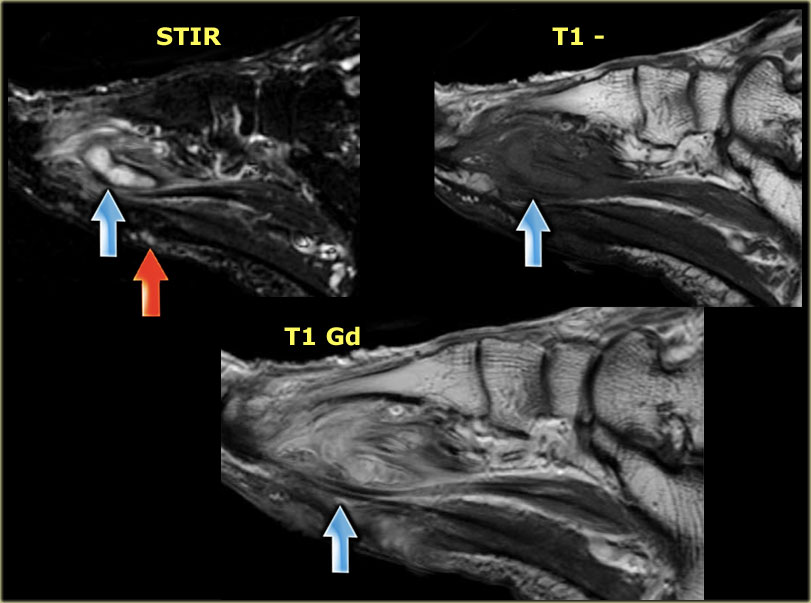
Lumbricals of foot are multiple small muscles that contribute biomechanical balance of the foot during walking. Head, neck, arm, foot, pelvis, etc. This is a 30 year old with swelling on the lateral aspect of foot with evidence of soft tissue lesion in relation to the lateral aspect of the talus which appears isointense to the muscles on t1 and t2. Learn about foot and ankle mri here. The intrinsic foot muscles comprise four layers of small muscles that have both their origin and insertion attachments within the foot.
Mri Of The Muscles In Wohlfart Kugelberg Welander Disease Journal Of The Neurological Sciences from els-jbs-prod-cdn.jbs.elsevierhealth.com Near normal foot mri for reference. Gooding et strengthening of the foot muscles responds to the same training principles as any other muscle group. Shop our pre workout and nitric oxide supplements. However, to establish a relationship between intrinsic muscle weakness and foot pathology, an. .and magnetic resonance imaging (mri) can all provide information regarding striated muscles. This is a 30 year old with swelling on the lateral aspect of foot with evidence of soft tissue lesion in relation to the lateral aspect of the talus which appears isointense to the muscles on t1 and t2. Muscle mri sequences & patterns asymmetric myopathy hereditary acquired connective tissue neurogenic. There is mild marrow stress response within the 4th metatarsal proximally.
Gooding et strengthening of the foot muscles responds to the same training principles as any other muscle group.
The flexor digiti minimi brevis (flexor brevis minimi digiti, flexor digiti quinti brevis) lies under the metatarsal bone on the little toe, and resembles one of the interossei. The foot is a complex structure whose functions are governed by numerous muscles, ligaments, tendons, nerves and joints that work together to provide balance and stability and produce movement. Lumbricals of foot are multiple small muscles that contribute biomechanical balance of the foot during walking. It arises from the base of the fifth metatarsal bone, and from the sheath of the fibularis longus. Mri with hardware in foot? This article reviews the use of magnetic resonance imaging (mri) in the evaluation of the foot, including a discussion of bone and cartilage abnormalities Case contributed by dr andrew dixon ◉. A quick reference guide describing the mrc muscle power assessment scale for neurological examination, including tables in various formats (including pdf). .magnetic resonance imaging (mri) or ultrasound imaging (usi) (soysa et al., 2012; Abdm, abductor digiti minimi muscle; Mri with hardware in foot? Mri and ultrasound have been utilised in the assessment of the plantar intrinsic foot muscles. Learn about foot and ankle mri here.
Near normal foot mri for reference. .magnetic resonance imaging (mri) or ultrasound imaging (usi) (soysa et al., 2012; Mri patterns of neuromuscular disease involvement thigh & other muscles 2. The flexor digiti minimi brevis (flexor brevis minimi digiti, flexor digiti quinti brevis) lies under the metatarsal bone on the little toe, and resembles one of the interossei. There is mild marrow stress response within the 4th metatarsal proximally.
Anatomy Of The Foot And Ankle Mri from www.imaios.com Mri with hardware in foot? Indications for foot mri scan. The foot is a complex structure whose functions are governed by numerous muscles, ligaments, tendons, nerves and joints that work together to provide balance and stability and produce movement. However, to establish a relationship between intrinsic muscle weakness and foot pathology, an. Learn more details about them at kenhub! Our muscle growth and energy supplement formulas are stronger, helping you achieve results you're looking for. Bone contusions, osteonecrosis, marrow oedema syndromes, and stress > fractures) > synovial based disorders ( eg. Magnetic resonance imaging (mri), with its multiplanar capabilities, superior soft tissue contrast, excellent spatial resolution, ability to image bone marrow, noninvasiveness, and lack…
Head, neck, arm, foot, pelvis, etc.
A quick reference guide describing the mrc muscle power assessment scale for neurological examination, including tables in various formats (including pdf). Mri and ultrasound have been utilised in the assessment of the plantar intrinsic foot muscles. Magnetic resonance imaging was not performed with the same mri scanner as used in the initial studies, but exactly reduced size of foot muscles using mri has been reported by greenman et al. Abdm, abductor digiti minimi muscle; Our muscle growth and energy supplement formulas are stronger, helping you achieve results you're looking for. Subscribe to foot & ankle problems. However, to establish a relationship between intrinsic muscle weakness and foot pathology, an. Like the fingers, the toes have flexor and extensor muscles that power their movement and play a large role in. Intrinsic foot muscle weakness has been implicated in a range of foot deformities and disorders. ► shoulder ► elbow ► wrist ► finger ► thumb. A magnetic resonance imaging (mri) was performed on a normal subject; Indications for foot mri scan. .magnetic resonance imaging (mri) or ultrasound imaging (usi) (soysa et al., 2012;